Table of Contents


Within the broader spectrum of dental procedures, perhaps no treatment suffers from a more enduring and unwarranted public relations crisis than root canal therapy. Historically, the phrase alone has evoked widespread clinical anxiety, often incorrectly associated with the cause of dental pain rather than its definitive cure. However, the field of endodontics—the specialized branch of dentistry focused on diagnosing and treating the internal tissues of the tooth—has undergone a profound technological renaissance over the last two decades. The transition from tactile, “blind” procedures to highly illuminated, microscopic precision has fundamentally redefined the success rates of endodontic therapy. Today, microscopic root canal treatment stands as a marvel of modern medical engineering, offering patients the most biologically sound alternative to tooth extraction and implant replacement.
The Pathogenesis of Pulpal Infection
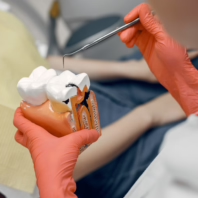
To understand the necessity of endodontic intervention, one must examine the complex internal architecture of human dentition. A tooth is not a solid block of enamel and dentin; its core houses a vital, highly vascularized network known as the dental pulp. This delicate tissue, extending from the crown down through the roots to the apex of the jawbone, contains the nerves, blood vessels, and connective tissues responsible for the tooth’s sensory functions and immunological defense.
When the protective outer layers of the tooth are breached—whether by deep cariogenic bacteria (severe decay), repeated restorative trauma, or a physical fracture—the sterile environment of the pulp chamber is compromised. Pathogenic oral bacteria invade the pulpal tissue, triggering a massive inflammatory immune response. Because the pulp is encased within rigid dentinal walls, the inflammatory swelling has nowhere to expand. This causes a dramatic increase in intrapulpal pressure, compressing the nerve fibers and resulting in the excruciating, throbbing pain classically associated with a “toothache.” If left untreated, the pulpal tissue undergoes necrosis (death), and the bacterial infection inevitably spreads through the tip of the root (the apex) into the surrounding alveolar bone, creating an apical abscess and threatening systemic health.
Talk to a Turkeyana consultant
Free evaluation and an all-inclusive quote within 24 hours.
The Paradigm Shift: From Tactile to Visual Dentistry
Historically, conventional root canal therapy was largely a tactile exercise. The clinician relied heavily on two-dimensional radiographs and the physical “feel” of hand files navigating the narrow root canal system. This blind approach inherently carried significant clinical risks, as human dental anatomy is rarely straightforward. Roots often curve dramatically, bifurcate into microscopic branches, or harbor hidden accessory canals.
The introduction of the Dental Operating Microscope (DOM) shattered these clinical limitations. A modern endodontic microscope provides up to 25x magnification combined with intense, coaxial, shadow-free illumination directed straight down the long axis of the tooth. This technological leap transitioned endodontics from a discipline of tactile guesswork to one of absolute visual precision. A prime example of this is the mesiobuccal 2 (MB2) canal, a microscopic fourth canal frequently present in upper first molars. Without a microscope, the MB2 is notoriously difficult to locate and is a leading cause of endodontic failure when missed. With high-powered magnification, the clinician can visually identify, meticulously clean, and completely seal these microscopic anatomical variations, exponentially increasing the long-term prognosis of the treated tooth.


Advanced Biomechanical Decontamination Protocols
Locating the complex canal network is only the first phase of the endodontic evolution; the subsequent decontamination process has also been entirely revolutionized. Traditional stainless-steel hand files have been largely replaced by engine-driven, nickel-titanium (NiTi) rotary instruments. NiTi alloys possess a property known as “shape memory” and extreme flexibility, allowing them to precisely follow the severe curvatures of a root canal without tearing or perforating the delicate dentinal walls.
Furthermore, the mechanical shaping of the canal is heavily supported by advanced chemical irrigation. Endodontists now utilize sophisticated ultrasonic devices to activate antimicrobial irrigants, such as sodium hypochlorite, within the canal. The acoustic streaming and cavitation bubbles generated by the ultrasonic energy propel the chemical disinfectants deep into the microscopic dentinal tubules and lateral canals that files cannot physically reach, ensuring the total eradication of the bacterial biofilm. Once the system is structurally prepared and biologically sterilized, it is hermetically sealed. Modern obturation techniques utilize warm vertical condensation of gutta-percha (a biocompatible rubber) paired with state-of-the-art bioceramic sealers. These sealers chemically bond to the dentin, expand slightly upon setting, and release calcium hydroxide, creating an impenetrable, bioactive barrier against future bacterial ingress.
The Biological Imperative of Preservation
In an era where dental implants are highly accessible, patients frequently question whether saving a severely infected tooth is worth the effort. From a strict biological and biomechanical perspective, the answer is unequivocally yes. The ultimate goal of dentistry is the preservation of natural human anatomy. A natural tooth is suspended within the jawbone by the periodontal ligament (PDL). This highly specialized connective tissue acts as a vital shock absorber and is rich in mechanoreceptors. These receptors provide proprioception—crucial sensory feedback to the brain regarding bite force and chewing pressure. Dental implants, which fuse directly to the bone, entirely lack this proprioceptive ability, occasionally leading to structural overload if a patient unconsciously bites down too hard. By saving the natural tooth through microscopic endodontics, the clinician preserves the PDL, maintains the natural contours of the gingival tissue, and retains the original structural integrity of the dental arch.
Navigating the complexities of advanced endodontic therapy requires entrusting one’s care to an institution that refuses to compromise on technological investment or clinical expertise. Turkeyana has established itself as a premier destination for international patients seeking to salvage their natural dentition rather than settling for immediate extraction. By equipping specialized endodontic suites with elite Dental Operating Microscopes, 3D Cone Beam Computed Tomography (CBCT) for precise anatomical mapping, and ultra-flexible rotary systems, the clinic ensures that even the most complex root canal anatomies are treated with absolute certainty. The highly accredited specialists at Turkeyana approach every infected tooth with a profound commitment to biomimetic preservation, transforming a historically feared procedure into a painless, highly predictable, and deeply restorative medical success. For those who understand that nothing truly replicates the brilliance of natural human biology, Turkeyana provides the definitive, world-class intervention required to secure the longevity of an authentic smile.
Your feedback helps us improve.
Thank you for your feedback.