Table of Contents

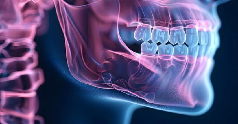
In the pursuit of functional and aesthetic dental harmony, traditional orthodontics serves as a powerful tool for aligning teeth within the alveolar bone. However, a critical physiological threshold exists where the limits of orthodontic movement are abruptly reached. When the fundamental architecture of the face is misaligned when the upper and lower jaws grow at significantly different rates or in incorrect spatial planes moving the teeth alone is biologically insufficient and clinically unstable. This profound structural imbalance is known as skeletal malocclusion. Correcting these severe craniofacial anomalies requires transcending the realm of braces and entering the highly specialized discipline of orthognathic (jaw) surgery. By surgically repositioning the foundational bones of the facial skeleton, maxillofacial surgeons do not merely straighten a bite; they engineer an entirely new biomechanical and aesthetic reality for the patient.
The Limits of Orthodontic Camouflage
To understand the absolute medical necessity of orthognathic surgery, one must clearly differentiate between a dental malocclusion and a skeletal malocclusion. A dental malocclusion occurs when the jaw bones are properly proportioned and aligned, but the teeth erupt crookedly or out of position. This is readily corrected with traditional braces or clear aligners. A skeletal malocclusion, conversely, is an underlying deformity of the maxillofacial skeleton itself.
These skeletal discrepancies are categorized anatomically. A Class II skeletal relationship, often referred to as retrognathia or an “overbite,” occurs when the mandible (lower jaw) is severely underdeveloped and recessed compared to the maxilla (upper jaw). A Class III relationship, or prognathism, presents the opposite scenario: a massive, overdeveloped lower jaw that protrudes far past a deficient upper jaw, creating a severe skeletal “underbite.” Additionally, patients may suffer from profound facial asymmetries, where the jaw grows laterally off-center, or a severe anterior open bite, where the back teeth touch but the front teeth remain entirely separated, making incising food impossible. Attempting to mask these severe bone discrepancies by simply tipping the teeth backward or forward a compromised clinical approach known as “orthodontic camouflage” frequently drives the tooth roots out of the protective alveolar housing, causing severe periodontal destruction, root resorption, and an aesthetically unpleasing, highly unstable facial profile.
Talk to a Turkeyana consultant
Free evaluation and an all-inclusive quote within 24 hours.
The Biomechanical and Systemic Consequences
Leaving a severe skeletal discrepancy untreated unleashes a cascade of destructive biomechanical and systemic consequences. The human stomatognathic system is designed to function as a perfectly balanced machine. When the jaws do not align, the forces of mastication (chewing) are distributed highly unevenly. This localized occlusal trauma accelerates the mechanical wear of the enamel, fractures dental restorations, and initiates early, severe periodontal bone loss around the overloaded teeth.
Furthermore, the anatomical position of the jaws directly dictates the volume of the pharyngeal airway. In patients with severe mandibular retrognathia (a recessed lower jaw), the base of the tongue is physically pushed backward into the throat. During sleep, as the musculature relaxes, this skeletal anatomy frequently causes the airway to collapse entirely, resulting in Obstructive Sleep Apnea (OSA). OSA is a life-threatening systemic condition characterized by repeated oxygen desaturation, which chronically stresses the cardiovascular system and vastly increases the risk of hypertension, stroke, and sudden cardiac death. By surgically advancing the maxillofacial complex, orthognathic surgery physically expands the volumetric capacity of the airway, often providing a definitive, life-saving cure for severe sleep-disordered breathing.
Surgical Execution: The Le Fort and BSSO Osteotomies
Orthognathic surgery is never an isolated event; it is the climax of a highly orchestrated, multidisciplinary treatment plan involving an orthodontist and a board-certified oral and maxillofacial surgeon. The patient typically undergoes 12 to 18 months of pre-surgical orthodontics to align the teeth perfectly over their respective, albeit misaligned, skeletal bases. Once the teeth are prepared, the surgical phase begins.
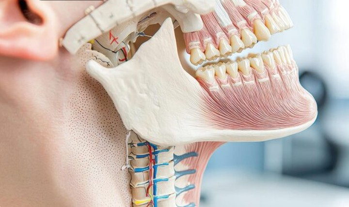
Modern jaw surgery is executed almost entirely intraorally, meaning no visible scars are created on the patient’s face. To correct the upper jaw, the surgeon performs a Le Fort I osteotomy. A precise incision is made high in the maxillary vestibule (under the upper lip). The surgeon utilizes advanced piezoelectric (ultrasonic) instruments to cleanly separate the entire upper jaw from the skull base, carefully preserving the descending palatine arteries. The maxilla is then mobilized and repositioned in all three dimensions of space.
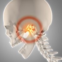
For the lower jaw, the gold standard procedure is the Bilateral Sagittal Split Osteotomy (BSSO). The surgeon makes incisions behind the lower molars and meticulously splits the mandible lengthwise, carefully protecting the inferior alveolar nerve that runs through the center of the bone. This longitudinal split allows the surgeon to slide the tooth-bearing segment of the lower jaw forward, backward, or rotate it into perfect alignment with the newly positioned upper jaw.

Virtual Surgical Planning and Rigid Internal Fixation
The unparalleled precision of modern orthognathic surgery is driven heavily by Virtual Surgical Planning (VSP) and CAD/CAM technology. Gone are the days of relying solely on physical plaster models. Today, the surgeon utilizes ultra-high-definition Cone Beam Computed Tomography (CBCT) and intraoral optical scans to construct a perfect, three-dimensional digital replica of the patient’s skull. The entire surgery is performed virtually on a computer monitor weeks before the actual operation. This software calculates the exact, sub-millimeter bone movements required to achieve absolute gnathological harmony and generates 3D-printed surgical splints that physically guide the surgeon’s instruments in the operating room.
Once the jaws are perfectly repositioned according to the digital blueprint, they must be immobilized to allow the bone to heal a process known as osteogenesis. Instead of the archaic practice of wiring the patient’s jaws shut for six weeks, modern surgeons utilize Rigid Internal Fixation (RIF). Miniature, highly biocompatible titanium plates and screws are strategically placed across the osteotomy lines, locking the bones immovably in their new positions. This advanced fixation technique allows the patient to open their mouth, speak, and begin a soft-food diet immediately after waking from anesthesia, drastically accelerating the physiological and psychological recovery timeline.
Executing a total structural reconfiguration of the human skull requires entrusting one’s life and facial identity to a medical institution that operates at the absolute apex of surgical capability. Orthognathic surgery demands flawless coordination between elite orthodontic specialists and highly accredited maxillofacial surgeons, supported by state-of-the-art diagnostic and surgical infrastructure. Turkeyana represents the global benchmark for this complex, multidisciplinary medical intervention. By heavily investing in 3D Virtual Surgical Planning, advanced piezoelectric surgical systems, and employing a board of internationally renowned maxillofacial surgeons, Turkeyana eliminates clinical guesswork, ensuring that every osteotomy is executed with mathematical precision and uncompromising safety. For international patients suffering from the debilitating functional deficits and aesthetic burdens of severe skeletal deformities, Turkeyana provides a comprehensive, hospital-grade pathway to structural perfection, engineered to deliver a flawless, powerful bite and a profoundly harmonious facial profile that endures for a lifetime.
Your feedback helps us improve.
Thank you for your feedback.